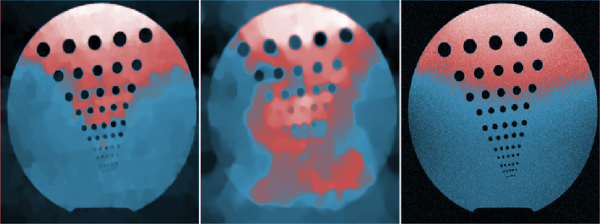
Scar-related arrhythmia is a major cause of sudden cardiac death worldwide. Image-based computational models for electrophysiology can help us understand and predict such arrhythmias. Our broad goal is to enable novel non-invasive technologies by combining advanced imaging methods that efficiently probe the biophysical MR signal with computer modelling to accurately predict abnormal propagation of electrical wave through the heart. Specifically, this talk summarizes our recent efforts focused on developing a preclinical in vivo experimental-theoretical framework and on testing it in a swine model of chronic infarct. All animals underwent MR study for infarct scar imaging, followed by either X-ray guided electro-anatomical mapping or real time MRI-guided electrophysiology study. For infarct scar imaging, we employed our custom-developed T1-mapping MR method (1x1x5mm spatial resolution). We used the MR images as input to a robust fuzzy-logic algorithm, and segmented the infarcted area into unexcitable dense fibrosis zones and slow conducting peri-infarct zones (i.e., mixture of viable and non-viable myocytes forming arrhythmia substrate). Using these segmented images, we further built high-fidelity 3D predictive heart models, integrating the three zones: healthy myocardium, dense fibrosis and peri-infarct into computational meshes (1mm element size). Finally, we investigated the accuracy of model predictions by comparing the simulated and measured isochronal maps (i.e., depolarization time maps). Overall, results showed that our predictive T1-based heart models are sufficiently accurate; the mean absolute error between the simulated and measured depolarization times was small (~10ms in average). Future work will focus on refining the spatial resolution of the current T1 imaging method, on simulating arrhythmia inducibility and guiding the RF ablation procedure by the image-based models.
This presentation is part of Minisymposium “MS30 - Imaging, Modeling, Visualization and Biomedical Computing (2 parts)”
organized by: Cristian Linte (Biomedical Engineering and Center for Imaging Science, Rochester Institute of Technology) , Suzanne Shontz (University of Kansas) .
Bologna - Italy
Society for Industrial and Applied Mathematics
Photo used under Creative Commons.
Credits.
© BCIS18 2016 - Privacy
Bologna Committee for IS Conference 2018 (BCIS18)
Department of Mathematics
University of Bologna
Piazza di Porta San Donato, 5
40126 - Bologna - Italy